Device and method for creating an optical tomogram of a living microscopic sample
Innovative Products and Technologies
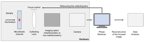
This technology describes a method and device for generating optical tomograms of microscopic samples using an advanced imaging process. The method involves rotating and displacing a sample within an optical microscope’s focal plane to capture a series of images from multiple angles. These images are subsequently processed to create a detailed three-dimensional model of the sample. The innovation reduces motion stress on living samples and minimizes the time required for tomogram acquisition. The device incorporates a rotatable sample stage and a precise imaging setup that enables high-resolution tomography suitable for applications in biological research, particularly on fragile, live samples.
Background
Conventional methods for 3D microscopic imaging, such as selective plane illumination microscopy (SPIM) and light sheet fluorescence microscopy (LSFM), are based on optical fluorescence and require labeled or stained specimens. Instead or in addition to the fluorescence imaging, it is advantageous to have alternative modalities using the same or similar sample mounting. However, some tomographic, high-resolution methods involving significant sample rotation are also restricted by mechanical limitations, which hinder efficient data acquisition and stress the sample. This innovation addresses these issues by introducing a smooth, continuous movement system that supports rapid image capture while maintaining minimal physical disruption to the specimen and that is compatible with existing light (sheet) microscopy techniques.
Technology
The device utilizes a microscope with a rotatable sample mounting, where a sample is rotated and simultaneously translated through the focal plane (Fig. 1A-B). This controlled movement forms a spiral imaging path that allows the sample to pass continuously through the focal plane at multiple angles, significantly reducing acquisition time. Typically, the spiral consists of 20 full rotations, images are acquired every 1° with high speed, e.g., at 60 frames per second (fps), and the total acquisition of 7200 images takes less than 2 min per specimen (Bassi et al., 2015). A bright-field or transmitted-light microscope captures digital images that are stored and processed to build a 3D model. This method’s innovative approach minimizes motion blur and mechanical stress by maintaining a continuous motion path, which enhances the stability of live sample imaging.
Advantages
Potential applications
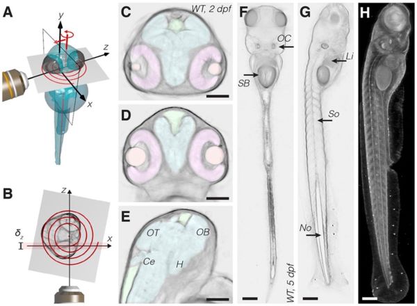
Figure 1:This figure illustrates the novel imaging technique for capturing high-resolution 3D images of live, transparent samples like zebrafish embryos. Panel (A) shows a schematic of the configuration, where the specimen is rotated smoothly through the focal plane while moving it along the detection axis, forming a spiral path. This movement allows multiple images from different angles to be captured without disturbing the sample. Panel (B) highlights how this spiral imaging creates a detailed, depth-resolved view. Panels (C) - (H) show reconstructed cross-sections of a zebrafish head and body, with clear views of organs such as retina (pink), eye lens (orange), brain ventricles (green), brain (cyan) (annotated brain domains: optic tectum (OT), hypothalamus (H), cerebellum (Ce) and olfactory bulb (OB)), swim bladder (SB), otic capsule (OC), liver (Li), somites (So), notochord (No). Scale bars: 100 µm. This technique provides a non-invasive way to visualize internal structures and follow development in live specimens over time (Bassi et al., 2015).
Patent Information
US10437038B2 (Application 20.02.2015)
Literature
Bassi et al., “Device and Method for creating an optical tomogram of a microscopic sample”, US 10,437,038B2, 8. October 2019
Bassi et al., Optical tomography complements light sheet microscopy for in toto imaging of zebrafish development. Development, 2015, 142. Jg., Nr. 5, S. 1016-1020.
Contact:
Dr. Bernd Ctortecka, M. Phil.
Senior Patent- & License Manager
Physicist
Email:
ctortecka@max-planck-innovation.de
As the central technology transfer company of the Max Planck Society, Max Planck Innovation has helped bridge the gap between science and industry since 1970.
The Max Planck Society (MPG) operates as Germany’s most successful organization in basic research and is worldrenowned for its cutting-edge research. In many cases this cutting-edge research also forms the basis for innovative products and services that are implemented through licensing and spin-off companies.
Max-Planck-Innovastion advises and supports scientists of the Max Planck Society in the assessment of inventions and filing of patents. We also market patents and technologies to industry, while coaching founders on how to build up new companies based on the research results of the Max Planck Society.
Create your free account to connect with Max-Planck-Innovation GmbH and thousands of other innovative organizations and professionals worldwide
Send a request for information
to Max-Planck-Innovation GmbH
Technology Offers on Innoget are directly posted
and managed by its members as well as evaluation of requests for information. Innoget is the trusted open innovation and science network aimed at directly connect industry needs with professionals online.
Need help requesting additional information or have questions regarding this Technology Offer?
Contact Innoget support