Whole Brain Visualization of Distinct Cortical Layers by MRI
Innovative Products and Technologies
An MRI platform including an imaging protocol and analysis software enabling in-vivo visualization, characterization and measurement of the distinct layers of the human cortex including width in 3D.
The imaging protocol utilizes inversion recovery (IR-MRI) pulse sequencing. A set of inversion times, identified and correlated to the different cortical layers provides the sought distinction between the layers.
The technology enables in-vivo research and diagnostics of abnormal morphology related to various diseases and disorders such as schizophrenia, autism, dyslexia, obsessive-compulsive disorder, Alzheimer's and mild cognitive impairment. Individual, clinical, comparative, quality and quantity information on the distinct layers at different developmental stages is made readily available. It also facilitates subject-specific brain segmentation for pre-surgical planning.
Project ID : 8-2013-443

The Technology
An MRI platform including an imaging protocol and analysis software enabling in-vivo visualization, characterization and measurement of the distinct layers of the human cortex including width in 3D.
The imaging protocol utilizes inversion recovery (IR-MRI) pulse sequencing. A set of inversion times, identified and correlated to the different cortical layers provides the sought distinction between the layers.
The technology enables in-vivo research and diagnostics of abnormal morphology related to various diseases and disorders such as schizophrenia, autism, dyslexia, obsessive-compulsive disorder, Alzheimer's and mild cognitive impairment. Individual, clinical, comparative, quality and quantity information on the distinct layers at different developmental stages is made readily available. It also facilitates subject-specific brain segmentation for pre-surgical planning.

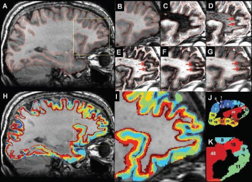
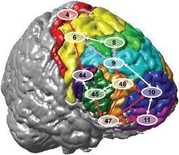
 An IR data set of the cortex. (A) A sagittal SPGR T1-weighted image of one representative slice with the cortex borders outlined in red. (B--G) Enlargement of the frontal part of cortex marked by the yellow box in (A) for the SPGR image (B): IR images with TI of 230 (C), 432 (D), 575 (E), 760 (F), and 1080 ms (G). The red arrows (D--F) show the propagation of the zero band of the IR along the cortex as TI increases.
 The Need
The cortex, termed also cortical gray matter, is morphologically composed of six cellular layers. These layers are the basis for separate neuro-anatomical regions referred to in "brain maps", routinely used in neuroimaging and neurosurgery. The width of these layers varies significantly between different brain regions, from 50 microns to 2 millimeters.
Conventional techniques for the characterization of cortical architecture are based on post-mortem observations. The presented technology introduces an ability to probe cortical layers (in correlation with the myelo and cyto architecture layers), in-vivo and non-invasively, providing a breakthrough for neurosciences.
Advantages
Project Status
Further development of 3D analysis capabilities is ongoing.
Patents
One patent application under examination.
Supporting Publications
Barazany D, Assaf Y. Visualization of Cortical Lamination Patterns with Magnetic Resonance Imaging. Cereb Cortex 2011